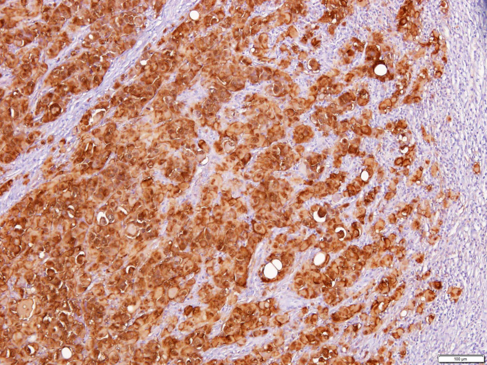
Rabbit anti-Human S-100, Polyclonal, Ig, Ready-to-use

Ready-to-use Antibody for Immunohistochemistry

SKU:
E031
Category
Quantity
6 ml

Background S-100

S-100 represents a group of acidic dimeric proteins that have a wide distribution in various tissues. Like calmodulin, parvalbumin, myoglobin, etc., they are calcium-binding and -modulating proteins. Three different dimers are known: S-100a (alpha/beta), S-100b (beta/beta) and S-100aa (alpha/alpha). The rabbit polyclonal antibody E031 reacts with human S-100a and S-100b. These proteins are typical of glial cells of the brain, ependyma, and Schwann cells of the central and peripheral nervous systems. S-100ao, on the other hand, is found in neurons, striated muscle cells, which usually do not stain or stain weakly with E031.

Synonyms

S-100, S-100a

Reagents sufficient for about 60-120 tissue sections

  • 1 dropper bottle HISTOPRIME® S-100 (Bottle, 6 ml)

Antigen

Purified S-100 protein from bovine brain

Specificity

S-100a and S-100b protein

Abnormal Tissue

Melanoma; ependymoma, glioma and schwannoma

Clone

Polyclonal

Isotype

Rabbit Ig

Fusion Partners

Rabbits were immunized with S-100 antigen isolated from ox brain. The antibody is present as a purified immunoglobulin fraction in stabilized buffer.

Among human central and peripheral nervous system tumors, S-100 positive reactions are found in schwannomas, ependymomas, and also in astrogliomas of various stages including glioblastomas. A large proportion of human tumor cells originating from salivary glands stain positively with S-100. Similarly, it can be detected in almost all nevus cell nevi and malignant melanomas. Langerhans cells in the epidermis react in the same way. In lymph nodes, interdigitating reticulum cells stain with E031.
The antibody E031 is directed against bovine S-100 and shows strong cross-reactivity with human protein; furthermore, reactivity with mouse, rat and chicken S-100 was observed.

Pretreatment

Proteolytic pretreatment with enzymes not required

Incubation Period

1 hour at room temperature

Usage

Ready-to-use in PBS, BSA, NaN3 (0,09%) pH 7,4(*) suitable on cryostat sections and on formalin-fixed, paraffin-embedded tissue sections.

Control Tissue

Skin, nerve tissue or appendix

Recommended Secondary Reagents

Alkaline Phosphatase Vectastain® ABC Rabbit IgG (Vector CatNo AK-5001) and SubstrateKit e.g. Vector® Red (Vector CatNo SK-5100).
Peroxidase Vectastain® ABC-Elite Rabbit IgG (Vector CatNo PK-6101) and Peroxidase Substrate-Kit e.g. DAB (LINARIS CatNo E108) or HistoGreen (LINARIS CatNo E109 ).

  • Moore, B.W. (1965). A soluble protein characteristic of the nervous system. Biochem. Biophys. Res. Comm. 19; 739-744.
  • Kindblom L.-G. Lodding P., Rosengren L., Baudier J., Haglid K. (1984) S-100 protein in melanocytic tumors. Acta Path. Microbiol.Immunol. Scand. Sect. A, 92:219.
  • Laurioal, L., Michetti, F., Sentinelli, S., Cocchia, D.: Detection of S-100 labelled cells in nasopharyngeal carcionoma. J. Clin. Pathol; 37: 12235-8 (1984).
  • Vanstapel M-J., Peeters B., Cordell J., Heyns W., De Wolf-Peeters Ch., Desmet V., and Mason D. (1985) Methods in Laboratory Investigation: Production of monoclonal antibodies directed against antigenic determinants common to the alpha- and beta- chain of bovine brain S-100 protein. Lab. Invest. 52; 232-238.
Specificity

S-100

Species Reactivity

Bovine, Cat, Horse, Human, Mouse, Rat, Swine

Host / Source

Rabbit

Isotype

Ig

Application

IHC-F, IHC-P

Clone

Polyclonal

Antigen

Human S-100

Quantity

6 ml

Format

RTU

Storage Temperature

2-8 °C

Shipping Temperature

20 °C

Documents

pdf Datasheet